Brachycephalic obstructive airway syndrome (boas) in Dogs and what we can do to help
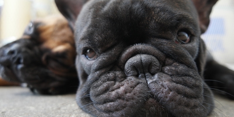
Brachycephalic Obstructive Airway Syndrome (BOAS) affects flat-faced (brachycephalic) dog breeds, such as French Bulldogs, English Bulldogs, Pugs and Boston Terriers.
These breeds are adored for their cute, squishy faces—but those same features can cause serious breathing problems.
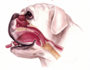
The term Brachycephalic obstructive airway Syndrome refers to the combination of:
Primary changes:
- Elongated and thick soft palate (tissue at the back of the throat which can protrude into the airways).
- Stenotic nares (narrow nostrils)
- Hypoplastic trachea (narrow windpipe)
- Abnormal nasal turbinates – abnormal cartilage in the back of the nose that restricts airflow.
Secondary changes:
- Everted laryngeal saccules (tissue that protrudes into the airway of the voice box)
- Enlarged tonsils and redundant pharyngeal tissue – contributes to airway obstruction.
The consequence of these challenges are:
- Noisy breathing that is worse during exercise or stress.
- Exercise intolerance and difficulty coping with hot temperatures
- Excessive snoring
- Choosing to sleep with their heads propped up or a toy held in their mouths
- Gagging or regurgitating, especially after eating or drinking
One way to think about these challenges is to imagine having to breathe through a “straw” in comparison to a “snorkel”, as the airways are much more narrow.
In the long term, the excessive effort required for breathing can lead to airway collapse, reduced quality of life and death. It also contributes to gastrointestinal issues due to increased negative pressure in the chest, and 97% of brachycephalic dogs will have oesophageal reflux issues, either going unnoticed, or causing clinical problems such as regurgitation – often seen as occasionally bringing up phlegm or exaggerated swallowing.
What can we do to help our pets?
Mild cases may be managed with weight control, limited exercise in heat, and medical therapy.
Moderate to severe cases often benefit greatly from surgical correction.. We perform 5/5 surgical correction in appropriate cases, in order to achieve the best outcome.
What is 5/5 surgical correction?
This refers to the comprehensive surgical correction of all five major BOAS abnormalities, when appropriate:
1. Nares Widening (Alarvestibuloplasty): Opens up the nostrils to improve airflow
2/3. H-phyaryngoplasty: Shortening and thinning of the soft palate to remove excessive tissue causing airway obstruction
4. Laryngeal Saccule Removal: Excises everted saccules if present, to reduce airway obstruction
5. Tonsillectomy: Removes enlarged tonsils if they obstruct airflow
The goal of surgery is to improve breathing efficiency, reduce airway pressure, and prevent long-term damage. Some physical issues such as narrow trachea cannot be corrected, but following surgery, breathing and gastrointestinal issues can be greatly improved.
What are the risks and how can we minimise these?
Airway surgery has the potential to improve a dogs quality of life and reduce serious life threatening respiratory events long term.
However, surgery in this delicate area can lead to some serious complications without proper care and attention before, during and after surgery. Possible complications include:
- Coughing
- Vomiting or regurgitation
- Aspiration (breathing in of fluid or food)
- Pneumonia (secondary to aspiration)
- Swelling of airways
- In rare cases, complications can lead to death
We minimise risks by taking the following measures:
- Use of pre-surgery medications to help reduce the risk of regurgitation and aspiration
- Medications to reduce anxiety if required
- A comprehensive and brachycephalic specific anaesthetic protocol, following the most up to date research and recommendations
- Intensive monitoring by highly trained nurses throughout the procedure and through recovery
- Use of anti-inflammatory medications to reduce risk of post operative swelling
- Introduce food in hospital 24 hours post operatively and monitor closely
- Feed soft food only in small meals for 2 weeks (ideally fed from hand)
- Use only harnesses for walking (no collars for 2 weeks minimum)
- Keep rested and confined at home for 2 weeks
- Monitor for cough and/or vomiting and treat as required
Complication rates are lower in young patients before signs of respiratory disease
In the ideal world, dogs would be monitored closely for the first 24-48 hours post operatively. Round the clock veterinary monitoring is available at specialist veterinary hospitals in Sydney and Canberra. Here at North Nowra Veterinary Hospital, our patients undergo surgery early in the day and are monitored closely until end of business at 6pm. At this time, dogs return home with their owners and are kept quiet and we ask owners to NOT offer food or water at this time. An after hours call service is available in cases of emergency.
The following day, patients return to hospital where they are offered small amounts of food and water and watched closely for normal swallowing and lack of coughing or vomiting. Weekly rechecks are recommended and after 2 weeks your dog can resume normal feeding and activity patterns.
At what age should surgery be performed?
BOAS surgery is best performed in young, healthy patients before significant secondary changes have developed. An ideal time is around 9-12 months of age, depending on breed, when dogs have mostly finished growing.
You may also like

Laparoscopic Desexing
Laparoscopic desexing offers an alternative method of desexing female dogs and cats from the traditional ovariohysterectomy. It is increasing in popularity amongst both veterinarians and pet owners as it can offer a number of significant benefits.

Laparoscopic Assisted Gastropexy in Dogs
What is GDV? GDV (gastric dilatation and volvulus) is a very sudden and serious condition seen in dogs where, due to range of factors, the stomach first dilates (bloats), and subsequently rotates (twists) on itself. This leads to the entrapment of fluid and gas in the...

Ear Infections
What is Otitis Externa and structure of the ear Otitis externa is inflammation of the external ear canal. The ear is divided into the outer, middle and inner ear. The outer ear includes the region from the ear flap (pinna) to the eardrum. The middle ear contains a...